也可用於 / Also available in:
 简体中文 (Chinese (Simplified))
Targeting, sizing, and phenotyping EVs from clinical samples is similar to finding a needle in a haystack. Nevertheless, flow cytometry is a practical solution and fast and can be done in a high throughput manner. To generate standardized and comparable EV flow cytometry results, reporting and calibration are of paramount importance for producing concordant results and discovering new clinical insights.
Parameters affecting EVs detection accuracy include background scattering caused by RI differences between the interface of the sample and sheath fluid, swarm detection, and autofluorescence signal generation, etc.
Researchers from the Vesicle Observation Center, Amsterdam University Medical Centers of The Netherlands have raised a few precautions when setting up a detection protocol for targeted EVs:
- Do not overlook the effect of EV composition and that composition property on the refractive index (RI). In the article, the measured scattering intensities of similar-sized beads of different materials (polystyrene (PS), silica, and hollow organosilica (HOBs)) of different RI is compared by a dedicated flow cytometer from Apogee Flow Systems, UK (model A60-Micro). In general, light scattering intensity increases with increasing diameter and RI. Using different beads from different materials for calibration will have different results.
- Using well-characterized reference beads for calibration so data can be converted to SI units and results from different systems and experiments can be compared directly as Mie theory can be used to relate the scatter signals to the diameter of EVs.
- The fluorescence intensity of labeled EVs can also be calibrated and compared by converting to molecules of the equivalent soluble fluorophore (MESF). In most cases, bright dyes such as APC and PE are recommended to have a better result.
To know more about the standardization of EV research, you may follow the METVES II project that focuses on new reference materials to standardize the flow rate, scattering, and fluorescence intensities of EV detection.
Reference:
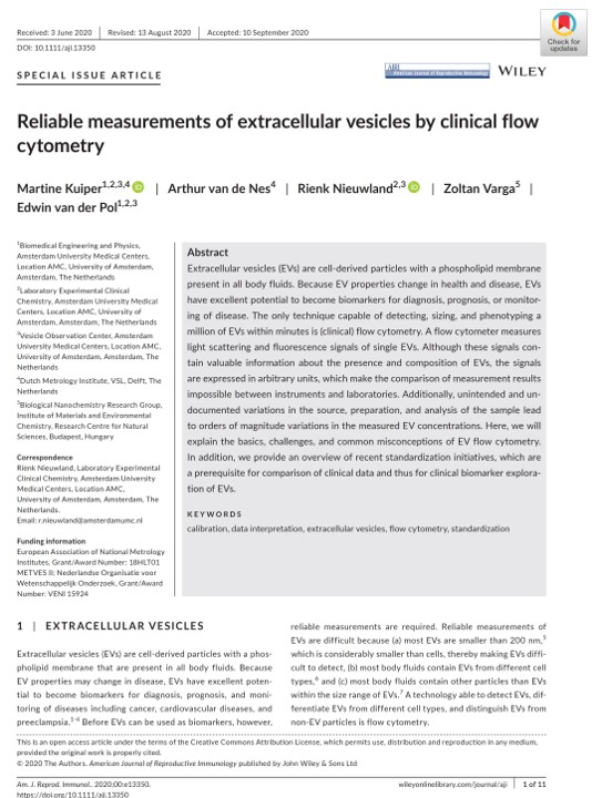
- Reliable measurements of extracellular vesicles by clinical flow cytometry. Kuiper M, et al. Am J Reprod Immunol. 2020. PMID: 32966654
- Publishable summary METVES II. Project Number: 18HLT01. Euramet. https://www.euramet.org/research-innovation/search-research-projects/details/project/standardisation-of-concentration-measurements-of-extracellular-vesicles-for-medical-diagnoses/. Published 2020, accessed May 18, 2020.


